
3D Microfluidic model for interstitial and intramural flows
Fluid flows in living systems can be broadly classified as intramural flows (flows within blood and lymphatic vessels) and interstitial flows (flows within tissues). Intramural flows cover a wide range of flow speed, from a few micrometers per second in lymphatic capillaries to tens of centimeters per second in arteries. Interstitial flows, driven by the hydrostatic and osmotic pressure differences between the arterial and venous, or arterial and lymphatic capillaries, are typically in the order of a few micrometers per second or Peclet number of about 1 at cell length scale. Interstitial flows have recently demonstrated to influence tumour cells directly via sheer stress, indirectly through matrix rearrangement by fibroblasts and by skewing the chemokine gradients in the ECM.
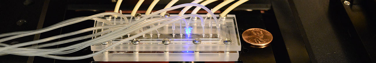
This work introduces a contact line pinning method to confine hydrogels such as natively derived collagen in a microfluidic channel without walls. By patterning collagen in designated wall-less channels, we demonstrated the generation of intramural flows through a microfluidic channel bounded by a mono-layer of endothelial cells (mimic of a vascular vessel), as well as slow interstitial flows within a cell embedded collagen matrix on the same microfluidic platform. The contact line pinning method ensures the generation of an engineered endothelial tube with straight walls, and spatially uniform interstitial fluid flows through the cell embedded 3D collagen matrix. Using this device, we demonstrated that the breast tumour cells' (MDA-MB-231 cell line) morphology and motility were modulated by the interstitial flows, and the motility of a sub-population of the cells was enhanced by the presence of the flow. The presented microfluidic platform provides a basic framework for studies of cellular behavior including cell transmigration, growth, and adhesion under well controlled interstitial and intramural flows, and within a physiologically realistic 3D co-culture setting.
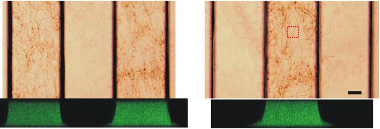
Confocal (side view) images of the three cell channels. Left image, collagen matrix is introduced into two side cell channels. This configuration is designed for generating an engineered endothelial tube for intramural flow studies. Right image, collagen is introduced in the middle cell channel. This configuration is designed for interstitial flow studies. Scale bar is 100 micrometer
Publications:
1. Chih-kuan Tung, Oleh Krupa, Elif Apyadin, Jr-Jiun Liou, Anthony Diaz-Santana, Beum Jun Kim, and Mingming Wu, A contact line pinning based microfluidic platform for modeling physiological flows, Lab-on-a-chip, 13(19):3876-3885 (2013).
Site created and maintained by Young Joon Suh